
Anemia is nearly ubiquitous in orthopaedic trauma patients. However, little is known about the impact of anemia and the associated iron deficiency on endochondral bone healing. We hypothesize that patients who receive IV iron infusions after orthopaedic trauma may have a different quantitative endochondral bone healing response compared to those who do not receive IV iron rescue. This research combines a current prospective randomized controlled trial examining the impact of IV iron infusions in orthopaedic trauma patients with ongoing research on collagen type X breakdown as a biomarker for endochondral bone healing. By quantifying the impact of IV iron rescue on bone healing, we hope to impact clinical practice and identify patients that may benefit from iron infusions following orthopaedic injuries. Additionally, this study will allow us to further characterize the systemic immune response after orthopaedic trauma and the impact of iron deficiency on that response.
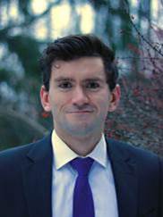
The OTA Resident Research Grant has provided an ideal opportunity to pursue clinical research grounded in basic science on a scale that can be accomplished while in residency. This project will allow me to contribute to orthopaedic knowledge as well as continue to grow my own skills as a researcher in ways that it would be difficult to do without this financial support. I'm very appreciative of the grant and look forward to using this opportunity to improve our understanding of the biologic processes underlying bone healing in trauma patients." - Catherine E T Hutchison, MD
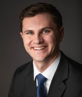
"The relationship between traumatic brain injury and fracture healing is poorly understood. Patients with traumatic brain injury have been shown to heal fractures faster and form more callus; however, the underlying mechanisms are largely unknown. Our project will examine the role of plasma leptin in accelerated fracture healing associated with traumatic brain injury. Radiographic healing and plasma leptin levels will be compared between patients with and without traumatic brain injury in the context of tibial or femoral shaft fractures. The findings of this study will shed light on the relationship between traumatic brain injury and fracture healing and may identify potential targets for novel therapeutic agents. I am very thankful for the OTA’s support through the Resident Research Grant. This grant will provide essential funding for our project." - Gareth Ryan, MD
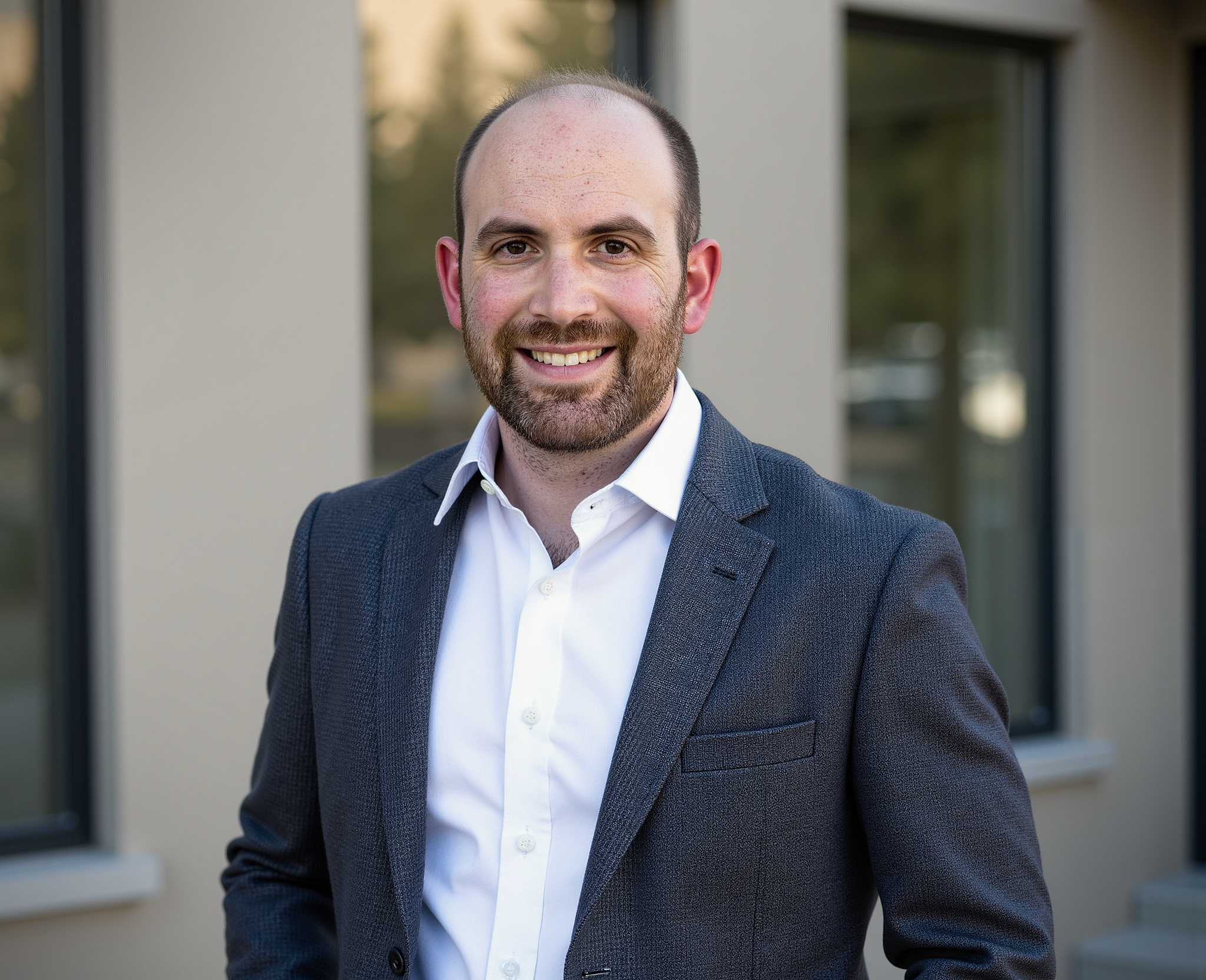
"Obtaining high-quality fluoroscopic images is a fundamental skill for all orthopedic residents that facilitates the safe placement of screws when treating pelvic and acetabular fractures percutaneously. Obtaining these views intraoperatively is technically challenging and requires a specific understanding of both fluoroscopy and pelvic anatomy. Nevertheless, obtaining and interpreting each view is considered a core competency for all orthopedic surgery residents and fellows. Generally, residents learn how to obtain and interpret these views through textbooks, lectures/videos, and teaching in the operating room. Occasionally, residents may have the opportunity to practice obtaining these views and placing percutaneous pelvic and acetabular screws in cadavers. However, there is currently no easily accessible and economic hands-on training model for residents to learn how to obtain and interpret these fluoroscopic images or to place percutaneous pelvic and acetabular screws safely and effectively. These concerns are particularly compounded at institutions with relatively lower cumulative trauma volumes and in resource-limited countries where access to cadavers and trauma fellowship training may be limited.
Therefore, the purpose of this study is to examine the effectiveness of a novel, affordable, realistic, and easily accessible pelvis training model and an interactive educational platform, known as “The Ortho Academy,” in teaching residents about obtaining and interpreting fluoroscopic images of the pelvis and acetabulum and performing percutaneous pelvic and acetabular wire/screw insertion. With validation, this teaching model can be easily incorporated into residency training and may even have a potential role for use in the Orthopaedic Trauma Association Resident Fracture Courses.
Our entire team is grateful to the OTA for supporting this research to advance education in this complex and ever-expanding area of orthopedics." - Nicholas Jerome Tucker, MD
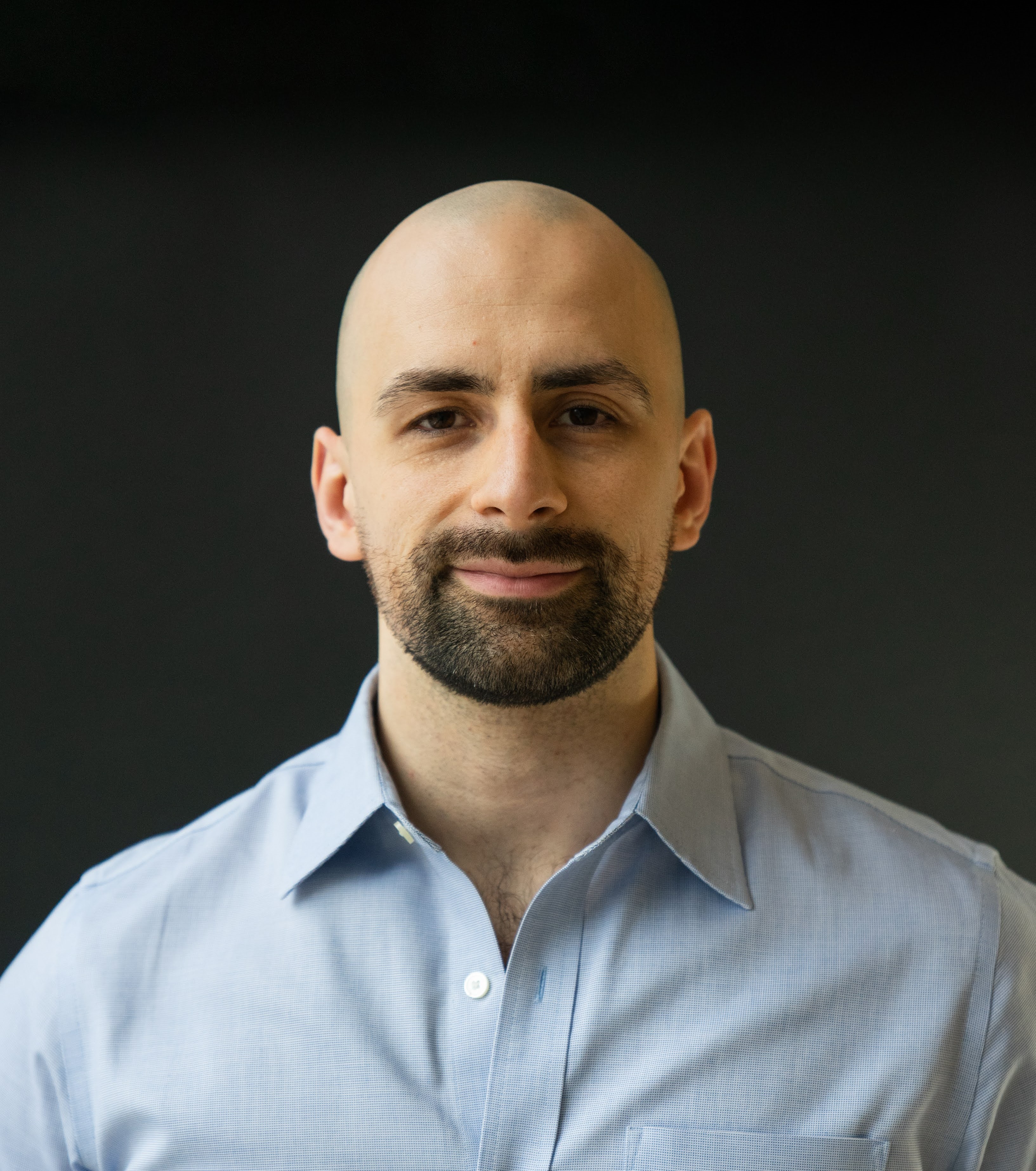
"After sustaining a lower extremity fracture, patients have a clear goal: recovery. Unfortunately, our current tools for measuring recovery—such as in-office examinations and patient-reported outcome measures (PROMs)—have inherent limitations, including subjectivity, measurement error, and recency bias. These assessments also provide only brief snapshots in time and lack comparison to a patient’s pre-injury baseline. Ideally, we would establish a passively collected, nonintrusive, longitudinal, and objective metric to better understand recovery trajectories and support personalized, patient-centered care.
Apple iPhones passively track mobility metrics such as step count, walking speed, double support time, stride length, and gait asymmetry—all of which have been independently validated. Our research team further validated these Apple Health metrics as reliable markers of recovery following lower extremity fractures and used them to develop individualized recovery curves. This work resulted in the publication of “Predicting Post-Fracture Recovery with Smartphone Mobility Data” in The Journal of Bone and Joint Surgery (JBJS), and a second manuscript, “The Future is Mobile: Pilot Validation Study of Apple Health Metrics in Orthopaedic Trauma,” has been accepted and is pending publication in JBJS.
I am incredibly grateful to the Orthopaedic Trauma Association for awarding me the Resident Research Grant, which provided the essential support to complete this project during residency. This work serves as the foundation for a planned multi-center cohort study and will inform future research across orthopaedic subspecialties." - Brian Shear, MD
"OTA Resident research grants have been instrumental in our lab’s work on developing, and now translating, a endothelial progenitor cell based approach to treat bone defects and non-unions. As many surgeons know, treating such disorders come with significant challenges. These injuries often require multiple, complex, and costly procedures to achieve union. At the same time, the current gold standard of autologous bone grafting provides an imperfect solution to this problem due to issues with donor site morbidity, graft volume, and overall effectiveness. The funding we have obtained from OTA has been key to both previous and current projects that have been a part of the development of this potential treatment for non-unions and bone defects which seeks to address these limitations and ultimately improve outcomes for patients suffering from these disorders. On a personal level, the OTA resident research grants have also been crucial in fostering my development as a budding academic trauma surgeon, having supported my Master’s thesis project on translating our promising preclinical findings, setting me on a trajectory to pursue similar work in the future. We are deeply appreciative of the OTA’s funding of our work, which has not only impacted myself and our labs work, but more importantly supports research that aims to have a direct impact on our patient’s and improve their outcomes." - Matthew Raleigh, MD
“Anemia is pervasive following orthopaedic injuries. Trauma and surgery contribute to acute blood loss as well as incite a systemic inflammatory response resulting in persistent injury-associated anemia even after adequate resuscitation and related functional iron deficiency despite normal iron stores. Previous work has begun to independently associate hemorrhagic shock and iron deficiency anemia with clinical nonunion and with delayed callus formation in preclinical models. The effect of injury-associated anemia on fracture healing remains unknown; our study is working to investigate these effects and the intervention of intravenous iron as a treatment in a controllable murine model.
This project would not be possible to complete as a resident without the OTA Resident Research Grant. The grant makes it possible for me to participate in translational research exploring the mechanism of this phenomenon in hopes that it may change the way we practice to better care for our patients. I am very appreciative of the experience as a principal investigator for this study as it has supported my development as I pursue my goal of becoming an academic surgeon-scientist.” - Natasha S McKibben, MD