Physical Therapy Videos - Foot & Ankle
What Is It?
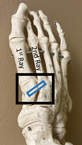
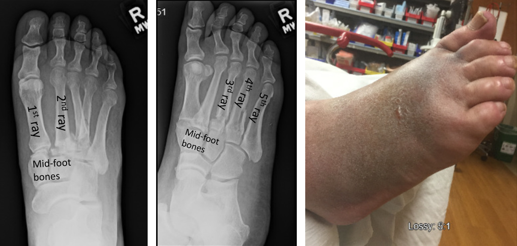
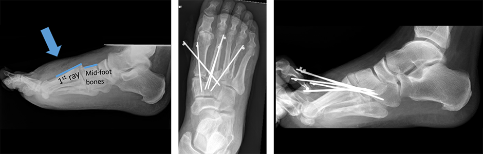
A "LisFranc" injury is an injury to the joint in the foot. It happens where long foot bones (metatarsals) meet the bones they connect to (tarsal bones). This injury can hurt the soft tissues (ligaments) and/or the bones. The injury can affect the whole foot or just part of it. Nerves and blood vessels near the bones can also be hurt. This injury can cause the foot to swell a lot. The bones can push on the skin, causing damage or blisters. Bones and ligaments are important for keeping your foot's shape. When you step down, they resist the force and keep the bones in their right places. When they're injured, your foot can collapse, causing pain while walking.
How It Happens
LisFranc injuries are rare and make up less than one percent of all fractures. They can happen after falling, a car accident, a crushing injury, or a sports injury. Different situations can cause different types of LisFranc injuries.
First Steps
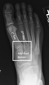
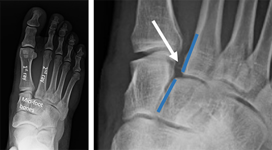
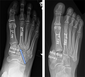
These injuries can cause pain and swelling in the middle of your foot when you try to walk or stand. A doctor will check your foot and take x-rays to see the injury. They might also take x-rays of your other foot for comparison. A CT scan might be needed to see more details of the injury. If needed, a doctor might try to push the bones back into place. A splint or boot can be used to protect your foot and help with the pain. You should avoid putting weight on your foot and keep it elevated to reduce swelling. You can go home and see an orthopedic surgeon later if your skin and bones are okay.
Treatment
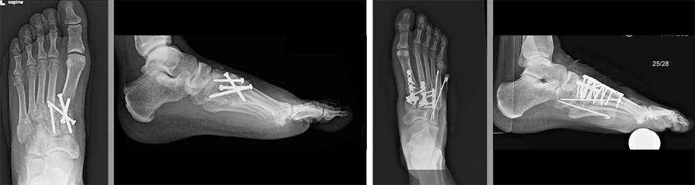
LisFranc injuries often need surgery. Sometimes, if the bones are lined up well, they can heal without surgery. In that case, a cast or splint is used and no weight is put on the foot for several weeks to months. Surgery can be done in different ways. Screws, plates, or pins can be used to hold the bones together. The surgeon will decide the best way to fix your injury.
Recovery
After surgery, it's important to keep your foot elevated to reduce swelling. You'll likely wear a splint, cast, or boot for several weeks to months. You won't be able to put weight on your foot for two to three months while it heals. You'll need to use crutches, a walker, a knee scooter, or a wheelchair to move around. Following your surgeon's instructions is important for proper healing.
Long Term
LisFranc injuries can have long-lasting effects on your foot. Some people can return to their normal activities, but others may have ongoing pain, stiffness, and weakness. This can happen even with successful surgery and healing. Your foot may not be as strong as before the injury. You might need to change your activities or wear special shoes or braces. Sometimes the screws or plates are taken out on purpose. This would need another surgery. Sometimes the bones don't heal and more surgery is needed to get the bones to heal.
Physical Therapy Videos - Foot & Ankle
More Information
---
Christopher Domes, MD
Edited by the OTA Patient Education Committee and David Sanders, MD (section lead)
All x-rays and pictures taken from the personal collection of Dr. Domes