Physical Therapy Videos - Femur
What Is It?
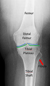
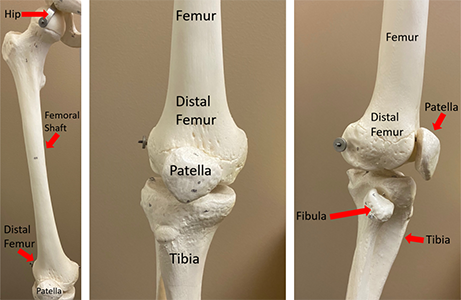
The lower part of your thigh bone is called the distal femur. It's shaped like a trapezoid and is found just behind your kneecap. Your knee is the biggest joint in your body that holds your weight. The end of the bone is covered with a smooth layer called cartilage, which cushions your knee and helps it bend. Strong muscles in the front and back of your knee and thigh help you bend and straighten your knee.
How It Happens
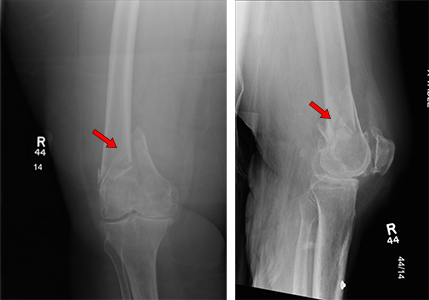
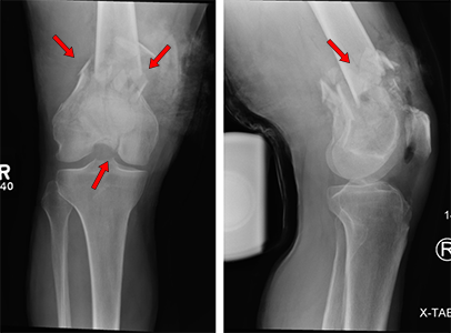
Distal femur fractures, or breaks in the lower part of your thigh bone, are not common. In young people, the bone is strong, so it takes a lot of force to break it, like in a car crash. Older people with weaker bones can break it from a simple fall. These fractures can affect your knee joint.
First Steps
These fractures are very painful when you try to move your leg or knee. You usually can't walk on it. A doctor will take x-rays and maybe a CT scan to see the break. They might put you in a knee brace to help with the pain and keep your leg stable. You'll probably be admitted to the hospital for more treatment.
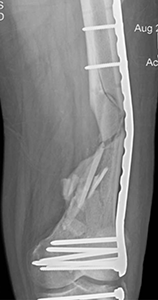
Treatment
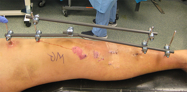
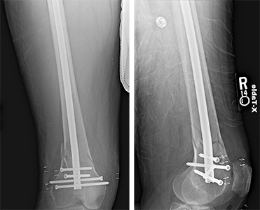
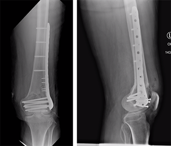
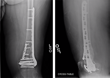
Most of the time, surgery is needed to treat distal femur fractures. It can take two to six months or more for the bone to heal. Surgery might happen one to five days after your injury, or it could be delayed if your leg is too swollen or you're not healthy enough. Sometimes, doctors use an "external fixator" (pins and bars outside your skin) to line up the bones and help with pain and movement until the swelling goes down enough for surgery. During surgery, the doctor makes cuts on the front, outside, or inside of your knee and thigh. They put the bones back together and use metal plates, screws, or a rod to hold them in place.
Recovery
While your leg is healing, you might not be able to put weight on it for six to twelve weeks. But some fractures are stable enough for weight-bearing right away. Your surgeon will decide. You can usually start moving your knee after surgery. Sometimes a special knee brace is used. Moving your ankle is important too, as it can get stiff. You may need a walker or cane when you start putting weight on your leg. Physical therapy can help you recover. Make sure to follow your doctor's instructions.
Long Term
Your injured leg might be weaker than your other leg for months. After surgery, you might have numbness around the scar and swelling in your leg. Swelling can last for months or never fully go away. The plates and screws can bother you, especially around the outside and inside of your knee. They might need to be removed after the fracture heals, which requires more surgery. If they don't bother you, there's usually no need to remove them. Long-term problems can include the break not healing, which can cause pain, weakness, and knee deformity. If the knee joint was injured this can cause arthritis. Arthritis usually causes pain and stiffness. If this happens, it's important to see your doctor, and you might need more surgery.
Physical Therapy Videos - Femur
More Information
---
Christopher Lee, MD
Edited by the OTA Patient Education Committee and Justin Haller, MD (section lead)
All x-rays and pictures taken from the personal collections of Dr. Lee and Christopher Domes, MD