Physical Therapy Videos - Elbow
What Is It?
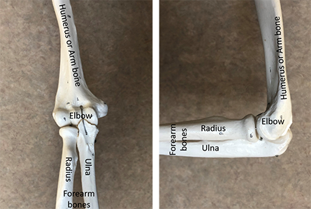
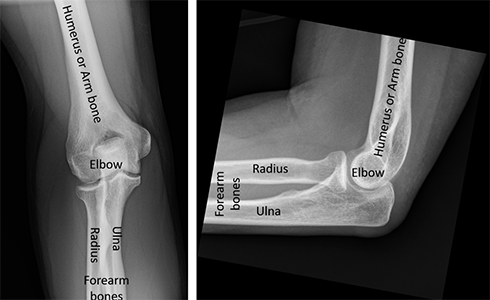
The elbow is the joint that connects your arm and forearm. It helps you move your hand closer to or away from your body. The elbow also lets you turn your hand palm-up and palm-down. Because the elbow is important for moving your hand, it's important to keep it working well after an injury.
How It Happens
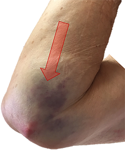
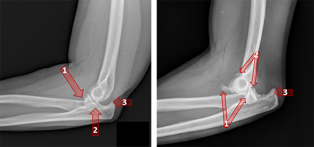
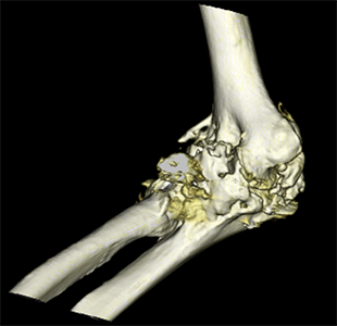
Elbow injuries can happen when you fall onto your outstretched hand. The bones in your forearm, the radius and ulna, might break. If the radius and ulna break at the elbow and the joint moves out of place, it's called a "terrible triad" injury. People with this injury have pain and can't move their arm well. They might also feel numbness or tingling in their hand. Sometimes, the blood supply to the arm is damaged.
First Steps
You need to go to a well-equipped emergency room for treatment. Doctors will check if the nerves and blood vessels in your arm are working. They will take x-rays to see if there are broken bones. If you have a dislocation, the doctors will try to put your elbow back in place. They might give you medicine to help with pain and make you sleepy. Your arm will be put in a splint, and you might have a CT scan to get a better look at your elbow. You might need surgery, depending on your injury.
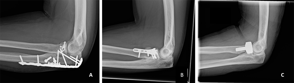
Treatment
Surgery is often needed for severe elbow injuries like this. If the elbow can be put back in place(reduced), surgery will happen after the swelling goes down. If the elbow can't be reduced, surgery will be sooner. During surgery, the doctor will make a cut over your elbow, fix the bones, and use metal plates and screws to hold them together. Sometimes, the top of the radius bone needs to be replaced with a metal piece. After surgery, your arm will be put in a splint, cast, or brace.
Recovery
After some days in a cast, splint, or brace, you will start moving your elbow. This helps you get back movement and stops your elbow from getting too stiff. Your doctor will limit your activities for six to twelve weeks after the injury. They might give you a special splint and tell you not to straighten your elbow all the way. This helps keep your elbow from dislocating again. Your doctor will check x-rays to make sure your elbow is still in the right place.
Long Term
After an elbow injury, you might have stiffness or dislocations again. It's common for people to not be able to straighten their arm all the way. But, you can still use your arm well even if it doesn't bend or straighten completely. Following your doctor's instructions and doing therapy can help. Other problems after surgery can include infections, irritation from metal parts, problems with bone healing, or extra bone forming. Sometimes, people with elbow injuries develop arthritis years later.
Physical Therapy Videos - Elbow
More Information
---
Christopher Doro, MD
Edited by the OTA Patient Education Committee and Steven Papp, MD (section lead)
All x-rays and pictures taken from the personal collection of Dr Doro