Physical Therapy Videos - Foot & Ankle
What Is It?
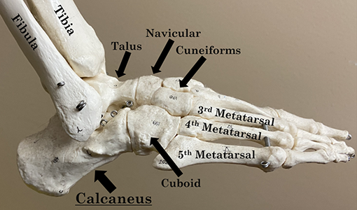
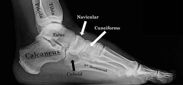
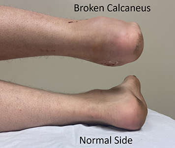
The calcaneus, or heel bone, is a large bone under your ankle and at the back of your foot. It helps you walk and connects your calf muscles to your foot, which lets you push off when you step forward.
How It Happens
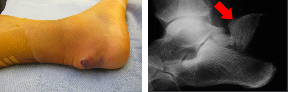
The calcaneus supports your body's weight, so it can get hurt if you fall from a ladder, roof, or have a car accident. The bone can break in different ways. If it breaks where the calf muscles connect, the broken pieces can move close to the skin, causing problems and maybe needing emergency surgery.
First Steps
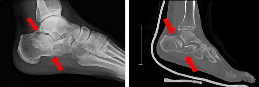
If you break your calcaneus, it usually hurts and swells right away. You might feel throbbing pain and see blisters. Your doctor will examine your foot and use x-rays or a CT scan to see the broken bone. They'll put your foot in a well-padded splint, boot, or partial cast to protect it. You should see a specialist or your regular doctor for follow-up care. Most people don't need to stay in the hospital, but sometimes urgent surgery is needed, and you may need to stay for that.
Treatment
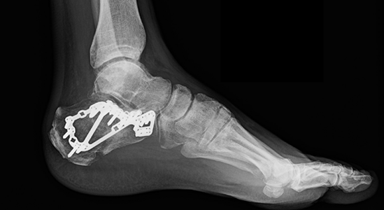
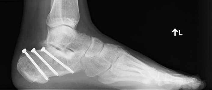
Some broken heel bones need surgery, but others don't. The bone takes about three to four months to heal either way. If you don't need surgery, you'll still need to limit movement and weight on the foot. Surgery might be needed if the break is inside a joint or if the bone's shape has changed. Talk to an orthopaedic surgeon about your injury and the best treatment. If the bone broke through the skin or is close to doing so, you'll almost always need surgery.
Recovery
While your heel bone heals, you won't be able to put weight on your foot. You might need physical therapy to help you move and put weight on the foot once it heals. It can take four to twelve weeks before you can put any weight on your foot, and even then, it might not be your full weight. Follow your surgeon's instructions for the best results.
Long Term
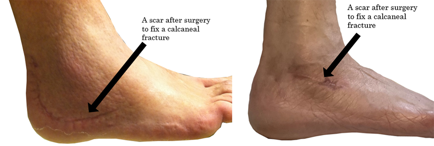
After a broken heel bone, you might have a limp or stiffness in your foot or ankle. Your heel could stay swollen compared to the other side. You might have pain when walking, especially on uneven ground. These problems usually get better over time, but your foot might always look or feel different. The joints in your injured foot can also get arthritis. If you had surgery, your surgeon will watch the wound to make sure it heals well. The metal plate and screws used to fix the bone are rarely noticed under the skin, but they might be removed later. In rare cases, if the bone doesn't heal or there's an infection, you might need more surgery. If you still have problems with your foot and heel after it heals, talk to your surgeon about whether more surgery could help.
Physical Therapy Videos - Foot & Ankle
More Information
---
Matthew D. Riedel, MD
Edited by Christopher Domes, MD and David W. Sanders, MD
Unless otherwise credited, all images taken from the personal collections of Dr. Riedel and Dr. Domes