Physical Therapy Videos - Foot & Ankle
What Is It?
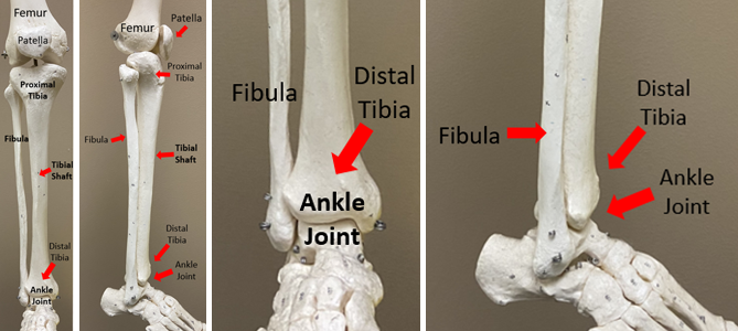
An ankle break, called a pilon fracture, is an injury to the ankle joint. The ankle is made of the tibia (shin bone), talus bone, and fibula (outside ankle bone). These bones have cartilage, a smooth surface that helps the ankle move easily. Ligaments connect the bones together. Blood vessels, nerves, and muscles are also part of the ankle.
How It Happens
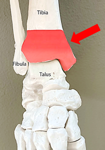
A pilon fracture is a break at the end of the shin bone. It can be a small crack or many broken pieces. This break usually happens after falls, car accidents, or work accidents. The talus bone pushes into the tibia and fibula, causing the break.
First Steps
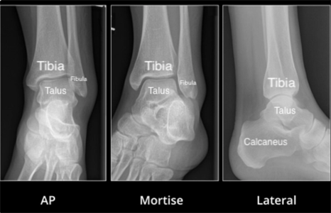
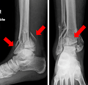
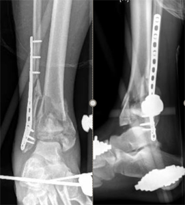
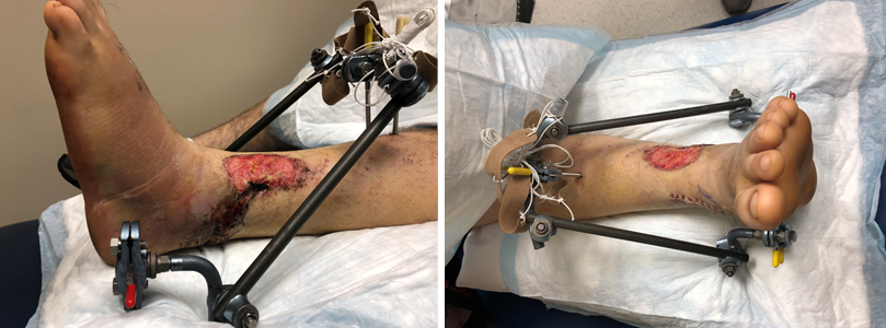
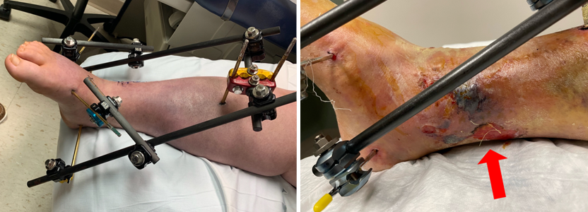
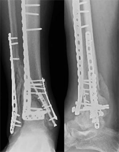
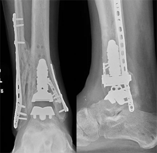
After a pilon fracture, it will hurt a lot and look crooked. You'll need to go to the emergency room. Doctors will take x-rays and maybe set the bones. They'll put your leg in a splint and you can't walk on it. You might need surgery to hold the bones in place. An external fixator may be used to hold the bones in place. This lets the swelling go down before a bigger surgery to fit the bones.
Treatment
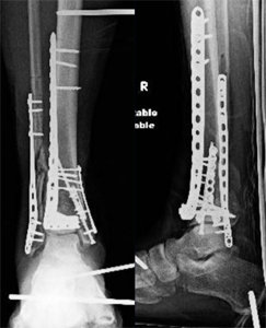
Your doctor will decide if you need surgery. It can take several cuts to fix the bones correctly. There needs to be minimal swelling in the ankle before this can be done. Your surgeon may use bone from a different area or a cadaver to replace your missing or destroyed bone. Most ankle breaks take 6 to 12 weeks to heal. After that, you'll slowly start walking more. You might need physical therapy to help with movement and walking.
Recovery
You can't put weight on your leg for weeks after the break. You might need crutches or a wheelchair. You'll wear a splint or brace for at least 2 weeks. After that, you may get a cast or another brace. Your rehab team might let you move your ankle soon after surgery. Be sure to follow your surgeon's instructions.
Long Term
Ankle breaks can cause arthritis, which might take years to develop. Patients might have stiffness, pain, and trouble walking. They might need braces or more surgery. An ankle break can cause long-term problems and pain. You might need more surgery or, in extreme cases, an amputation. If you stand or walk a lot at work, an ankle break will affect your life. You may need a job change or help with walking and standing.
Physical Therapy Videos - Foot & Ankle
More Information
---
Eric Magnuson, MD, and Christopher Domes, MD
Edited by the OTA Patient Education Committee
X-rays and images from the personal collections of Dr. Magnuson, Dr. Domes, and Matthew Hogue, MD