Physical Therapy Videos - Foot & Ankle
What Is It?
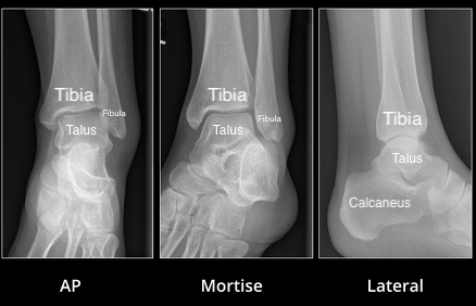
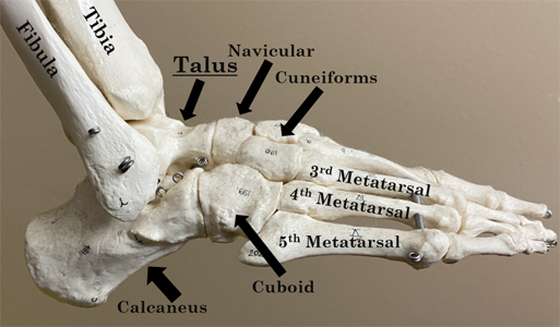
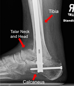
The talus is a bone in your foot that helps make up the ankle joint. It is between your shin bone (tibia) and heel bone (calcaneus). The joint between the talus and calcaneus is called the subtalar joint. This joint helps you move your foot side to side and is important for walking on uneven ground. The talus gets blood from small arteries. If this bone breaks, it can hurt the blood supply, which affects healing. Strong bands called ligaments hold the talus in place. These can also get hurt and make the talus unstable.
How It Happens
The talus is a strong bone because it holds up your whole body weight. To break it, you need a lot of force, like falling off a ladder or a car accident. The most common place it breaks is the middle part, called the neck. The neck is between the body of the talus (by the ankle joint) and the head of the talus (by the foot).
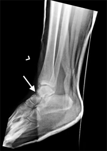
Another common place for the talus to break is on the outside part of the bone, called the "lateral process." This can happen when the foot is forced outward. It's sometimes called a "snowboarder fracture" because it happens a lot when snowboarding. In more serious injuries, the talus can break and move out of place (dislocate) at the ankle joint, the subtalar joint, or both. When this happens, a doctor needs to put the bones and joints back in place quickly.

Treatment
Talus fractures usually hurt a lot. The first step is to put your foot in a splint to keep it from moving, which can help with the pain. You might also need a CT scan to help your surgeon decide if you need surgery and if any other foot bones are broken. Most of the time, you need surgery for a talus fracture. But if the broken bone is in a good position and is stable, you might be treated with just a splint or cast. If the bones are far out of place, you may need surgery right away. The bones may need to be held in place with an external fixator or pins until a bigger surgery can be done
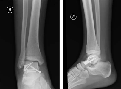
In rare cases, some or all of the talus might be missing or broken so badly it will not heal. If that happens, your surgeon may fuse the remaining bone with screws, long rods (nails), or plates and screws.
Recovery
After surgery, you'll be in a splint or cast to help your bones and soft tissues heal. Usually, you can't put any weight on your foot. If you put too much weight on it before the bones heal, the fracture might move. It takes about eight to twelve weeks for the bone to heal. As you get better, your doctor will take more x-rays to see how your bones are healing.
Long Term
Talus fractures can be hard to recover from. Some people have pain, stiffness, and swelling even after the bones heal. This can get better over time, but you might always notice a difference compared to your other foot. Because talus fractures often happen in high-energy events, like falling from a high place, the soft part of the joint (cartilage) can get hurt. This can lead to arthritis in the joint. If it gets really bad, you might need more surgery like a joint fusion or ankle replacement. Over time, your surgeon will want more x-rays to check the position of the fracture and the bone's health. If the blood supply to the bone was damaged, the bone cells might die. This is called avascular necrosis, and it can happen after a talus fracture. If it does, you might need more surgery.
Physical Therapy Videos - Foot & Ankle
More Information
---
Ashlee MacDonald, MD and Gillian Soles, MD
Edited by the OTA Patient Education Committee
X rays and images from the personal collection of Dr. MacDonald, Dr. Soles and Christopher Domes, MD