Physical Therapy Videos - Shoulder
What Is It?
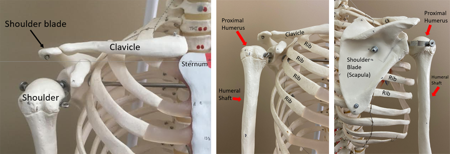
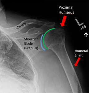
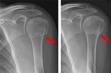
Your shoulder has three bones: the collarbone, shoulder blade, and upper arm bone. The top part of the upper arm bone is called the proximal part. Many muscles are around your shoulder. Blood vessels and nerves go from under your collarbone down your arm.
How It Happens
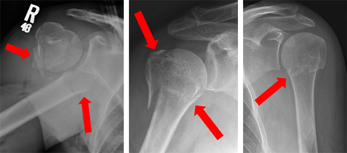
A broken shoulder is common in older people. It can happen after a simple fall. In younger people, it takes more force, like a car crash or a fall from a height. There are many ways the bone can break. You should talk to your doctor about your specific break.
Treatment
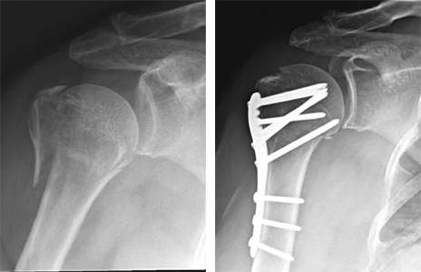
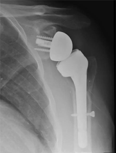
Most broken shoulders can be treated without surgery. It takes three to four months for the bone to heal. During this time, you will do exercises to get better movement and strength. Sometimes, surgery is needed. A surgeon will talk to you about the best choice for your injury. If you need surgery, the doctor will fix your bone with metal plates and screws. If your shoulder is in many pieces, the doctor might recommend a shoulder replacement instead.
Recovery
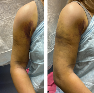
While your shoulder is healing, you might not be able to move it fully or lift heavy things. You will do physical therapy to help with movement and pain. A sling might be used for comfort. It's important to move your elbow, wrist, and hand to avoid stiffness. Follow your surgeon's instructions for the best results.
Long Term
After a broken shoulder, you might have stiffness, soreness, or not be as strong as before. You might need months of physical therapy. Your shoulder might always feel and move differently. Some people might have problems with the bone not healing or arthritis. These problems can happen with or without surgery. In older people, a shoulder replacement might be needed. In younger people, the doctor might suggest other surgery options.
Physical Therapy Videos - Shoulder
More Information
Adults:
Children:
---
Babar Shafiq, MD
Edited by the OTA Patient Education Committee and Steven Papp, MD (section lead)
X-rays and images from the personal collections of Dr. Shafiq and Christopher Domes, MD