Session VII - Pediatrics
Effect of Interposed Periosteum in an Animal Physeal Fracture Model
Laura Senunas Phieffer, MD; J. Michael Wattenbarger, MD; Ralph A. Meyer, Jr., PhD; Helen E. Gruber, PhD; Mark Easley, MD, Carolinas Medical Center, Charlotte, NC
Purpose: Many orthopaedic surgeons believe that interposed periosteum limiting the reduction of a physeal fracture should be removed since it may lead to a physeal bar. The purpose of this study was to observe the effect of interposed periosteum on physeal fracture healing in an animal model.
Materials and Methods: Fifty-two skeletally immature female Harlan Sprague-Dawley rats, age 4-5 weeks, were randomized to one of three cohort groups. All animals underwent surgical dissection of the left proximal medial tibia. Group I (fracture alone) underwent physeal fracture; Group II (fracture + periosteum) underwent physeal fracture and interposition of periosteum in the fracture site; Group III (positive control) underwent physeal fracture, excision of half of the growth plate, with interposition of periosteum in the defect. Fractures were induced by manually applying a valgus force. Initially, external fixation was used in all animals. Weekly radiographs were evaluated (blindly) for physeal bar formation and leg-length discrepancy (LLD). Leg-length discrepancies were calculated comparing right (control) versus left (experimental) tibia. Animals were euthanized on postoperative days (POD) 21 and 84. After harvest, tibia were fixed, decalcified, embedded in paraffin, serially sectioned in the coronal plane, and stained with Masson's trichrome. Histology specimens were evaluated for evidence of physeal bar formation and physeal morphology. Standard statistical methods were performed. P< 0.05 was considered significant.
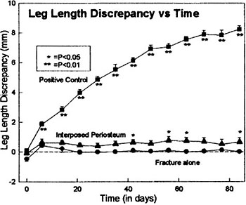
Results: All animals in Group III (positive control) showed radiographic bar formation by POD 14 with LLD averaging 8.23+0.26 (mean+SEM) mm by POD 84. In contrast, Group I's (fracture alone) LLD was 0.02+0.09 mm, and Group II's (fracture + periosteum) LLD was 0.67+0.28 mm by POD 84 respectively. There was a statistical difference in LLD between all groups, although the absolute difference was small between group I and II. No animals in Group I revealed radiographic evidence of bar formation. Two animals in group II, one POD 21 animal and one POD 84 animal, showed radiographic evidence of bar formation with LLDs of 1.2 mm and 2.6 mm respectively, at time of euthanasia. A significant difference in LLD was found when comparing Group I vs. II at POD 21 and 84 (P<0.05). In all Group III animals, at POD 21, histologic analysis revealed large areas of physeal arrest (47+3% of physeal width). Four out of 8 animals in Group I, and 5 out of 8 animals in Group II developed smaller bars (6+3% and 10+3% respectively) by POD 21. No significant differences in bar size, or percent and severity of physeal disorganization were found between Groups I and II, but significant differences were found when comparing both Groups I and II vs. III.
Discussion: Fractures involving the growth plate are common in children and are usually treated by nonoperative methods. Accepted orthopaedic dogma has been that interposed periosteum limiting reduction of a physeal fracture should be removed from the fracture site because it may lead to physeal growth arrest. Despite the accepted orthopaedic practice of operative intervention in suspected periosteal interposition, a comprehensive literature search identified no studies substantiating that the presence of periosteum enfolded in a physeal fracture has adverse effects on physeal fracture healing. Varying degrees of physeal bar formation occurred as a result of the physeal fracture alone without appearing to affect leg length. Interposition of the periosteum resulted in a small percent of animals with greater propensity of physeal growth disturbances, with resultant leg-length discrepancies, than fracture alone. Progressive leg-length discrepancies with extensive bar formation were present in all of Group III (positive controls) animals. Interestingly, Group I and Group II animals with smaller bars did not appear to behave differently than those without localization of bars across the physis.
Conclusions: We have developed a physeal fracture model in the proximal tibial metaphysis of skeletally immature rats. In this study, we found a small but significant difference in leg-length discrepancies between Group II (fracture + periosteum) and Group I (fracture alone).
Acknowledgements: We are greatly indebted to Odette Kangey, Leslie Sell and Audrey Stasky for their assistance with this project.