Thurs., 10/13/11 Basic Science, Paper #36, 4:41 pm OTA-2011
A Biomechanical Analysis of Lag Screw Position in the Femoral Head for Cephalomedullary Nails
Paul R.T. Kuzyk, MASc, MD, FRCS(C)1; Rad Zdero, PhD2,3; Suraj Shah, MEng Cand.2,3;
Michael Olsen, PhD2; James P. Waddell, MD, FRCS(C)1; Emil H. Schemitsch, MD, FRCS(C)1,2;
1Div. of Orthopaedics, Dept. of Surgery, University of Toronto, Toronto, Ontario, Canada;
2Martin Orthopaedic Biomechanics Lab., St. Michael’s Hospital, Toronto, Ontario, Canada;
3Dept. of Mechanical and Industrial Engineering, Ryerson University, Toronto, Ontario, Canada
Purpose: The purpose of this study was to determine if lag screw position affects the biomechanical properties of a cephalomedullary nail used to fix an unstable peritrochanteric fracture.
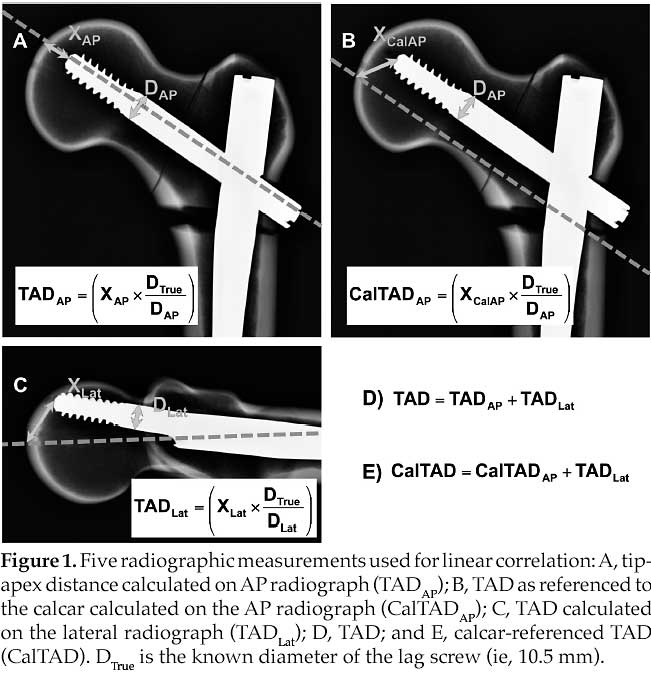
Methods: Unstable peritrochanteric fractures were created in 30 synthetic femurs and repaired with long Gamma3 nails using one of 5 lag screw positions: superior, inferior, anterior, posterior, central. Radiographic measurements including tip-apex distance (TAD) and a calcar-referenced TAD (CalTAD) were calculated from AP and lateral radiographs (Figure 1). Specimens were tested for axial, lateral, and torsional stiffness, and then loaded to failure in the axial position. Analysis of variance and linear regression were used for statistical analysis.

Results: The inferior lag screw position had significantly greater mean axial stiffness than superior (P <0.01), anterior (P = 0.02), and posterior (P = 0.04) positions. Analysis revealed significantly less mean torsional stiffness for the superior lag screw position compared to other lag screw positions (P <0.01 all 4 pairings). No statistically significant differences were noted for lateral stiffness. Superior and central lag screw positions had significantly greater mean load to failure than anterior (P <0.01 and P = 0.02) and posterior (P <0.01 and P = 0.05) positions. There were significant negative linear correlations between stiffness with distance from the calcar on AP radiographs, and load to failure with distance from the center of femoral neck on the lateral radiographs.
Conclusions: The inferior lag screw position produced the highest axial and torsional stiffness. Anterior and posterior lag screw positions produced the lowest stiffness and load to failure. For a cephalomedullary nail, in contrast to a sliding hip screw, we therefore suggest inferior placement of the lag screw, just above the calcar, on the AP radiograph and central placement in the femoral neck on the lateral radiograph, such that CalTAD is minimized.
Alphabetical Disclosure Listing (628K PDF)
• The FDA has not cleared this drug and/or medical device for the use described in this presentation (i.e., the drug or medical device is being discussed for an “off label” use). ◆FDA information not available at time of printing. Δ OTA Grant.