Thurs., 10/14/10 Basic Science, Paper #38, 4:46 pm OTA-2010
Altered Bone Quality in Bisphosphonate-Related Femoral Fractures of Postmenopausal Women
Brian J. Rebolledo, BA1; Eve Donnelly, PhD2; Dean G. Lorich, MD2; Brian P. Gladnick, BA1;
Aasis Unnanuntana, MD2; Adele L. Boskey, PhD2; Joseph M. Lane, MD2;
1Weill Cornell Medical College, New York, New York, USA;
2Hospital for Special Surgery, New York, New York, USA
Purpose: Bisphosphonates have proven to be useful in the prevention of bone loss in osteoporotic patients, yet less is known about the effects on bone quality. Recent reports have proposed that long-term bisphosphonate treatment (BIS+) could adversely affect bone quality by resulting in accumulation of microdamage and leading to low-energy “atypical” transverse fractures of the femoral diaphysis. In this study, we compared the microarchitectural and compositional parameters of bone quality in femoral fracture patients on BIS+ versus bisphosphonates-naive (BIS–) patients.
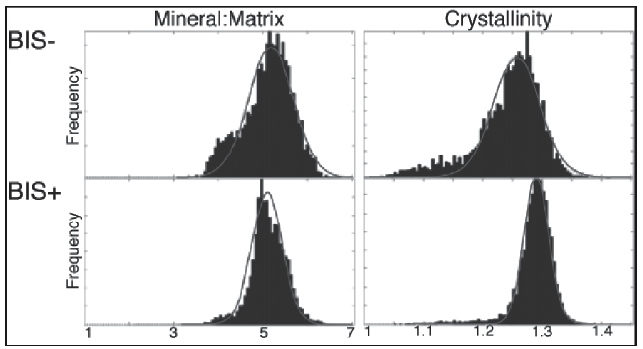
Methods: 21 postmenopausal women with proximal femoral fractures requiring internal fixation and trochanteric femoral nail (TFN) had corticocancellous bone biopsies obtained from the lateral proximal femur, distal to the greater trochanter. In the BIS+ group, there were 12 patients (age 81 ± 12 years) with average treatment duration of 8.5 ± 5 years. There were 9 patients (age 86 ± 6 years) in the BIS– group. Specimens were first analyzed by micro CT to measure bone volume fraction, trabecular (Tb) number, Tb thickness, Tb separation, and Tb connectivity. Histomorphometry was used to measure the ratio of unmineralized to mineralized bone surface of cortical and Tb bone. Specimens were then analyzed with Fourier transform infrared imaging (FTIRI) to assess properties of cortical and Tb bone with the following parameters: mineral:matrix ratio, carbonate:phosphate ratio, collagen cross-linking maturity, and mineral crystallinity. Each distribution was measured by mean values and full width at half maximum (FWHM) of the Gaussian curve fit to the distribution. Groups were compared with the Student t test, with P < 0.05 being significant.
Results: Cortical tissue mineral properties indicated a narrower distribution in the BIS+ group compared to the BIS– group (mineral:matrix ratio: –23%, P = 0.03; mineral crystallinity: –36%, P = 0.02) (Fig. 1).
While the widths of distribution values were different between groups, the mean cortical and trabecular values of all FTIRI parameters were similar. Micro CT and histomorphometry analysis showed no difference between groups. There was no difference in demographic data between groups.
Conclusion: In this study, BIS+ in femoral fracture patients was associated with a narrower distribution in compositional parameters of cortical tissue. The primary abnormality was a narrower pattern of aged bone matrix. Older unremodeled collagen can undergo glycation and result in reduced toughness. Narrow tissue distributions of bone have been reported previously in nonfractured osteoporotic patients on BIS+ for up to 3 years, and may be associated with a loss of mechanical integrity. This may also contribute to difficulties in preparing the femoral shaft for TFN and cause delay in the healing process. Using FTIRI, these preliminary results suggest narrower tissue properties detected in patients on longterm BIS+ could lead to adverse effects on bone quality and increase the risk of fracture.
Alphabetical Disclosure Listing (292K PDF)
• The FDA has not cleared this drug and/or medical device for the use described in this presentation (i.e., the drug or medical device is being discussed for an “off label” use). ◆FDA information not available at time of printing. Δ OTA Grant.