Session II - Pelvis & Acetabulum
Thurs., 10/8/09 Pelvis & Acetabulum, Paper #33, 4:10 pm OTA-2009
Nonoperative Immediate Weightbearing of Minimally Displaced Lateral Compression Sacral Fractures Does Not Result in Displacement
Gillian Sembler, MD (n); John Lien, MD (n);
Paul Tornetta, III, MD (3, 5A, 7-Smith &Nephew; 8-Exploramed);
Boston University Medical Center, Boston, Massachusetts, USA
Purpose: Lateral compression sacral fractures are the most common type of pelvic ring fracture. Although many surgeons employ nonoperative treatment, some surgeons advocate operative fixation to prevent deformity. No large series of minimally displaced fractures treated with immediate weightbearing has been published. The purpose of this study is to compare the initial and follow-up radiographs of patients with minimally (<10 mm) displaced lateral compression sacral fractures (LC1) treated nonoperatively with immediate weightbearing to determine the amount of displacement that occurs during healing.
Methods: We evaluated 120 patients who presented to a single Level 1 trauma center with a unilateral lateral compression sacral fracture with <10 mm of displacement, and had complete radiographs through healing. There were 70 women and 50 men whose average age was 46 years and ISS score was 15 ± 11. Sacral fractures demonstrating >1 cm of displacement or that were not lateral compression in nature were excluded. Nonoperative treatment consisted of immediate foot-flat mobilization and advancement of weightbearing as tolerated. Repeat radiographs were routinely obtained once the patient had ambulated 50 feet, or at 1 week to look for further displacement. Patients were followed with AP radiographs in the clinic at the 4- to 6-week and 10- to 12-week periods, and then every 6 to 8 weeks until they were healed. Specific measurements were made on the initial and follow-up radiographs by 2 independent observers not involved in the treatment of the patients. A vertical plumb line drawn through the center of the S1 and S2 bodies served as an anchoring point for measurements. Key landmarks were measured on each side of the pelvis, including the superior border of the iliac wing, superior border of the sacral body, the inferior border of the ischial tuberosity, as well as sacral body width. These specific measurements allowed for determination of the initial and final displacements, as well as the change in displacements during healing.
Results: The average displacement of the injured side upon presentation and the shift that occurred during union is seen in the table below. Of note, 27 of the iliac wing heights, 14 of the sacral heights, and 22 of the ischial heights were negative, meaning that on the AP radiograph, they were inferior to the uninjured side in the plane of the plumb line. The reported displacements are in absolute values so that negative displacements do not mask the amount of displacement.
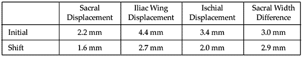
The greatest displacement over time was seen in the height of the ischium, with a maximum of 11 mm of change without a similar change in the height of the posterior ring measurements, possibly indicating flexion displacement. One patient failed nonoperative management after 1 week, demonstrating 5 mm of additional sacral displacement and having substantial pain with attempts to mobilize. She was treated with closed reduction and percutaneous pinning of the sacrum and an anterior external fixator. The other 119 patients (99%) mobilized well, healed with minimal additional displacement, and did not require surgical intervention.
Conclusions: We treated all patients presenting to our center with lateral compression sacral fractures having <10 mm of displacement (LC1 injuries) nonoperatively. One patient of the 120 displaced further and had clinical symptoms of pain warranting surgical intervention, while the remaining 119 healed without substantial further displacement. Immediate weightbearing, tempered by patient comfort, is a safe and acceptable treatment for minimally displaced lateral compression sacral fractures and results in union without substantial further displacement. Surgical management is not required to prevent deformity.
Disclosure: (n=Respondent answered 'No' to all items indicating no conflicts; 1=Board member/owner/officer/committee appointments; 2=Medical/Orthopaedic Publications; 3=Royalties; 4=Speakers bureau/paid presentations; 5A=Paid consultant or employee; 5B=Unpaid consultant; 6=Research or institutional support from a publisher; 7=Research or institutional support from a company or supplier; 8=Stock or Stock Options; 9=Other financial/material support from a publisher; 10=Other financial/material support from a company or supplier).
• The FDA has not cleared this drug and/or medical device for the use described in this presentation (i.e., the drug or medical device is being discussed for an “off label” use). ◆FDA information not available at time of printing. Δ OTA Grant