Thurs., 10/16/08 Tibia/Polytrauma, Paper #7, 3:43 pm OTA-2008
Tibial Metaphyseal Fractures: Nailing in Extension
Paul Tornetta, III, MD (c-Lippincott; a,c,e-Smith + Nephew); Brandon Steen, MD (n); Scott Ryan, MD (n);
Boston University Medical Center, Boston, Massachusetts, USA
Introduction: Fractures extending into the proximal and distal metaphyseal regions of the tibia are difficult to treat with intramedullary nails. Nailing in extension allows for easier visualization of the fracture, use of percutaneous clamps and blocking screws, and removes the flexion moment on proximal fractures. However, the use of a portal above the patella and the partial arthrotomy needed raises concerns for knee pain. The purpose of this report is to review a large series of metaphyseal fractures treated with nailing in extension using a superior portal, and to compare knee pain with a group of patients treated with a standard inferior incision.
Methods: Over a 7-year period, a single surgeon treated tibial fractures affecting the metaphysis by nailing with the knee in relative extension. A 3- to 5-cm incision was made from 1 cm above the patella extending distally. A partial superior medial arthrotomy allowed for subluxation of the patella. Special smooth and long jigs were used to avoid injury to the trochlear groove. A cannula system and alignment grid has been recently added. All patients were followed at regular intervals and a standardized knee pain (patient-based) score recorded at each visit. Patients with diaphyseal fractures treated by the same surgeon during the same period using a 1-cm infrapatellar incision in hyperflexion were used as a comparison group. Knee pain was graded on a 0 to 3 scale as per Court-Brown.
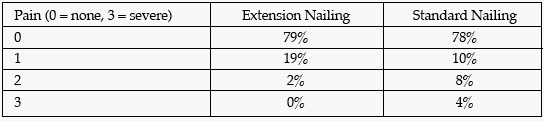
Results: 192 patients (148 male; 44 female) ages 16 to 87 years (mean, 40.1) with 68 open and 124 closed tibia fractures were treated with intramedullary nails. 85 had fractures extending into the proximal (66) or distal (19) metaphysis and were nailed in extension. The remaining 107 shaft fractures nailed in standard fashion comprised the control group. All nails were statically locked with multiplanar proximal screws. 17 proximal and 9 distal fractures extended into the joint. In 15 of the proximal and 6 distal fractures, lag screws and/or buttress plates were used to stabilize the joint prior to nailing. Radiographic angulation at the fracture was <3° in all patients, and no patient had malrotation of >5° by clinical examination. No patient lost reduction. Knee motion was within 10° of the normal side in all but two of the patients nailed in extension, both of whom had significant intra-articular injuries and obtained 110° and 120° of motion. For the healed fractures, knee pain was not different (P = 0.89) for the groups at an average follow-up of 48 weeks (range, 20-118 weeks) (see table).
Discussion and Conclusion: In this series, we report the use of extension nailing for both proximal and distal fractures extending into the metaphysis. This approach permits excellent biplanar imaging for clamp and blocking screw placement and, in contradistinction to earlier work, there were no significant angulatory defomities in these proximal and distal fractures nor any losses of reduction. Further, concerns regarding nailing through the knee were not borne out by a comparison with a group of patients nailed via a 1-cm incision in hyperflexion by the same surgeon over the same time period. We recommend this technique for tibia fractures extending into the metaphysis.
If noted, the author indicates something of value received. The codes are identified as a-research or institutional support; b-miscellaneous funding; c-royalties; d-stock options; e-consultant or employee; n-no conflicts disclosed, and *disclosure not available at time of printing.
• The FDA has not cleared this drug and/or medical device for the use described in this presentation (i.e., the drug or medical device is being discussed for an “off label” use). ◆FDA information not available at time of printing. Δ OTA Grant.