OTA 1997 Posters - Scientific Basis for Fracture Care
The Vacuum Sealing Technique - A Clinical Trial
Thomas Muellner, MD, L. Mrkonjic, O. Kwasny, V. Vécsei
Univ. Clinic of Traumatology, Vienna, Austria
The aim of this study was to prospectively evaluate the efficacy of a vacuum sealing technique (VST) in acute traumatic soft tissue defects and infected soft tissue defects complicated by exposed bone and/or implants as current treatment options are costly, traumatic, and occasionally ineffective.
Twelve patients sustained acute soft tissue defects at the knee or lower leg with a mean size of 22 sq cm (6-80 sq cm). Sixteen patients developed an infected soft tissue defect involving exposed bone and/or implants following rigid stabilization of a lower extremity fracture. Seven were closed and 9 were open fractures (3 grade I and 6 grade III). Initially the soft tissue defects averaged 12 sq cm (8-18 sq cm). All 16 patients had exposed bone with a mean area of 8 sq cm (6-10 sq cm). Six patients also had exposed implants with a mean area of 2.8 sq cm (1-4 sq cm). No implants were removed prior to fracture healing.
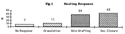
Twenty-six patients responded to the VST (Fig 1). Wound healing was achieved either by granulation, secondary closure or by split-thickness skin grafting. Active osteomyelitis was present in both cases in which the VST failed.
Polyvinyl foam under negative pressure generates an area of high contact forces at the wound-foam interface. This situation appears to facilitate granulation tissue production while maintaining a relatively clean wound bed. In 93% (26/28) of the patients the use of the VST following irrigation and debridement decreased the dimensions of the initial wound thus facilitating healing time and the eradication of any pre-existing infection. Wound closure by granulation, secondary closure, or split-thickness skin grafting was achieved in 26 wounds. The vacuum sealing technique is an effective option in the management of infected wounds.
|